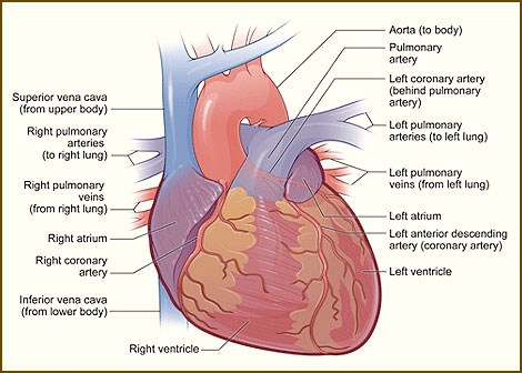
How the Heart Works – Exterior View
Anatomy of the Heart
Your heart is located under the ribcage in the center of your chest between your right and left lung. It’s shaped like an upside-down pear. Its muscular walls beat, or contract, pumping blood continuously to all parts of your body. The size of your heart can vary depending on your age, size, or the condition of your heart. A normal, healthy, adult heart most often is the size of an average clenched adult fist. Some diseases of the heart can cause it to become larger.
The Exterior of the Heart
Below is a picture of the outside of a normal, healthy, human heart.

The illustration shows the front surface of the heart, including the coronary arteries and major blood vessels. The heart is the muscle in the lower half of the picture. The heart has four chambers. The right and left atria (AY-tree-uh) are shown in purple. The right and left ventricles (VEN-trih-kuls) are shown in red. Connected to the heart are some of the main blood vessels—arteries and veins—that make up your blood circulatory system. The ventricle on the right side of your heart pumps blood from the heart to your lungs. When you breathe air in, oxygen passes from your lungs through blood vessels where it’s added to your blood. Carbon dioxide, a waste product, is passed from your blood through blood vessels to your lungs and is removed from your body when you breathe air out.
The atrium on the left side of your heart receives oxygen-rich blood from the lungs. The pumping action of your left ventricle sends this oxygen-rich blood through the aorta (a main artery) to the rest of your body.
The Right Side of Your Heart
The superior and inferior vena cavae are in blue to the left of the muscle as you look at the picture. These veins are the largest veins in your body. They carry used (oxygen-poor) blood to the right atrium of your heart. “Used” blood has had its oxygen removed and used by your body’s organs and tissues. The superior vena cava carries used blood from the upper parts of your body, including your head, chest, arms, and neck. The inferior vena cava carries used blood from the lower parts of your body. The used blood from the vena cavae flows into your heart’s right atrium and then on to the right ventricle. From the right ventricle, the used blood is pumped through the pulmonary (PULL-mun-ary) arteries (in blue in the center of picture) to your lungs. Here, through many small, thin blood vessels called capillaries, your blood picks up oxygen needed by all the areas of your body. The oxygen-rich blood passes from your lungs back to your heart through the pulmonary veins (in red to the left of the right atrium in the picture).
The Left Side of Your Heart
Oxygen-rich blood from your lungs passes through the pulmonary veins (in red to the right of the left atrium in the picture). It enters the left atrium and is pumped into the left ventricle. From the left ventricle, your blood is pumped to the rest of your body through the aorta. Like all of your organs, your heart needs blood rich with oxygen. This oxygen is supplied through the coronary arteries as it’s pumped out of your heart’s left ventricle. Your coronary arteries are located on your heart’s surface at the beginning of the aorta. Your coronary arteries (shown in red in the drawing) carry oxygen-rich blood to all parts of your heart.


